
This page contains specialized information intended exclusively for authorized medical professionals. By continuing, you confirm that you are a physician or medical provider.
Injectable MACT
Injectable MACT (Matrix-Associated Autologous Chondrocyte Transplantation) builds on more than 20 years of clinical experience with autologous chondrocyte transplantation (ACT) and combines autologous chondrocytes with a hydrogel matrix. The cell-hydrogel product is injected arthroscopically directly into the cartilage defect and does not require additional fixation. In clinical studies and earlier publications, this procedure has also been referred to as hydrogel-based ACT.
.avif)
Indication
For symptomatic focal cartilage defects of the knee (ICRS Grade III-IV) with defect sizes ≥2 cm² [1]
Fully Arthroscopic Application
In appropriate patients, the cell-hydrogel product can be applied arthroscopically using a dual-chamber syringe [3]. No trimming of the transplant required, no fixation necessary [2].
In Situ Cross-Linking Hydrogel
The hydrogel forms a stable matrix in situ within 1-3 minutes and retains the cells within the defect without the need for additional fixation [1, 2].
Understanding the Procedure
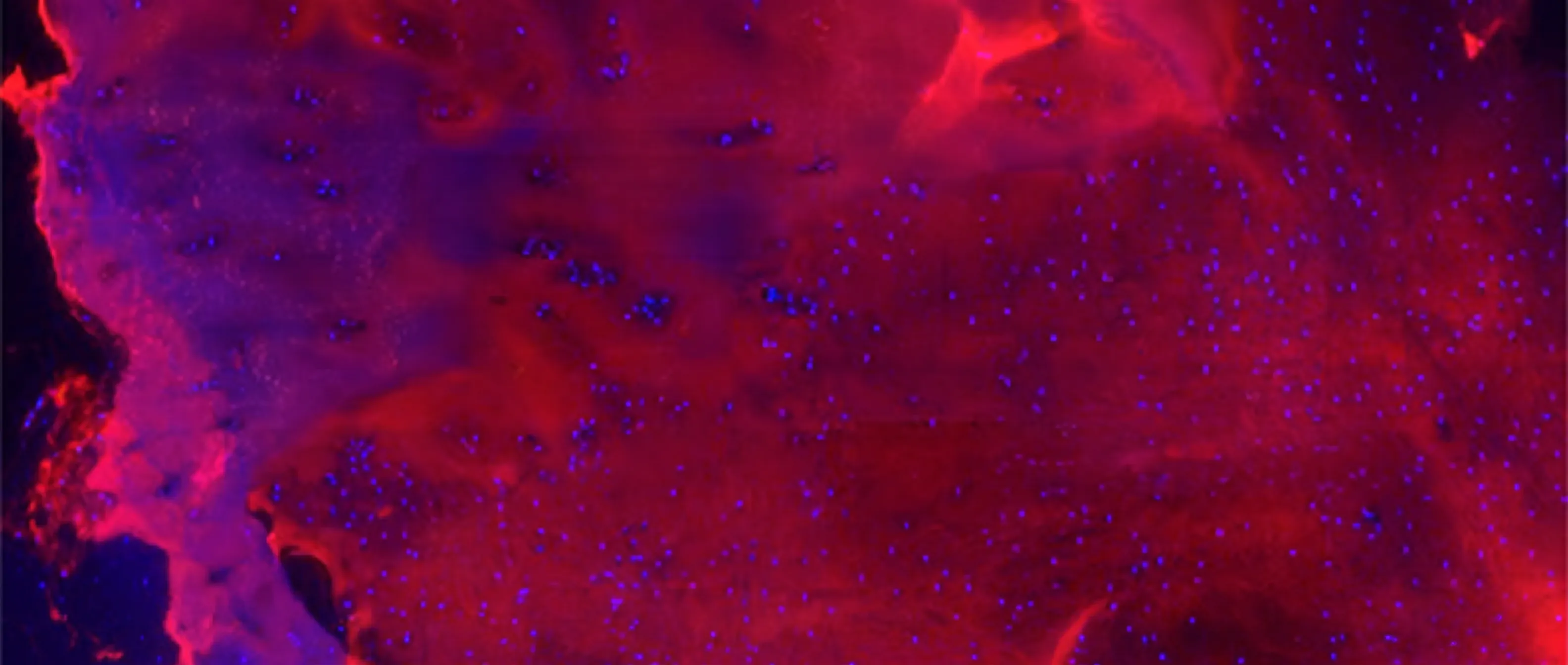
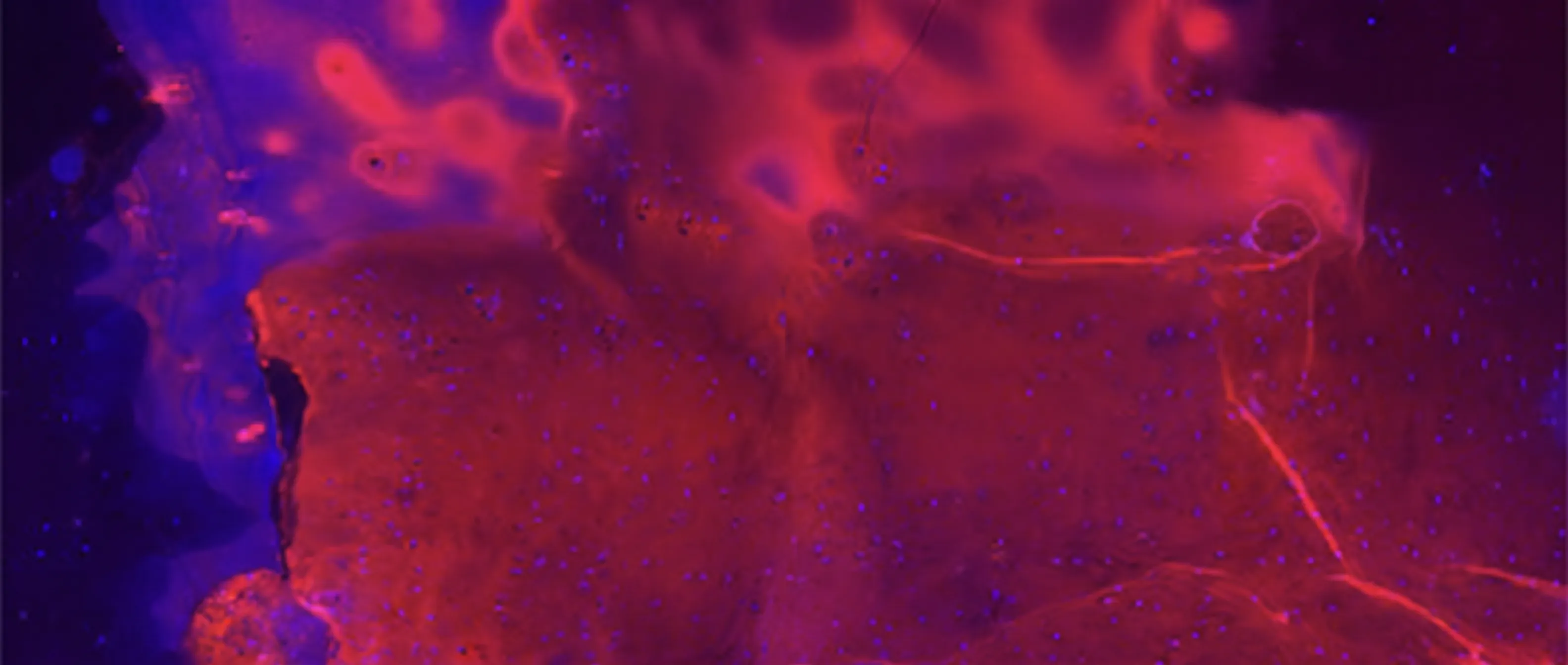
In Injectable MACT, following harvest of small osteochondral biopsies, autologous chondrocytes are expanded ex vivo and subsequently injected into the cartilage defect together with a hydrogel using a dual-chamber syringe. The hydrogel cross-links in situ and retains the transplanted cells within the defect, eliminating the need for additional fixation or defect coverage. Arthroscopic delivery also allows treatment of anatomically difficult-to-access defect locations, including retropatellar defects. The transplanted cells support cartilage regeneration. Imaging and histological evaluations have indicated the formation of repair tissue with hyaline properties.
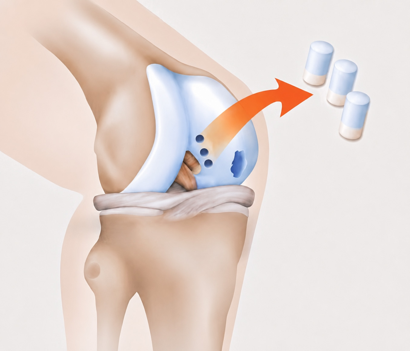
Tissue Harvest
Performed arthroscopically through standard portals
3 cartilage-bone cylinders are harvested from a non-load-bearing site (e.g., intercondylar notch)
Tissue is sent to TETEC for transplant manufacturing
.jpg)
Transplant Manufacturing
Autologous cells isolated from the biopsies are expanded ex vivo and combined with a hydrogel component. For implantation, a crosslinking agent is provided separately and co-administered with the cell product, enabling in situ gel formation at the defect site.
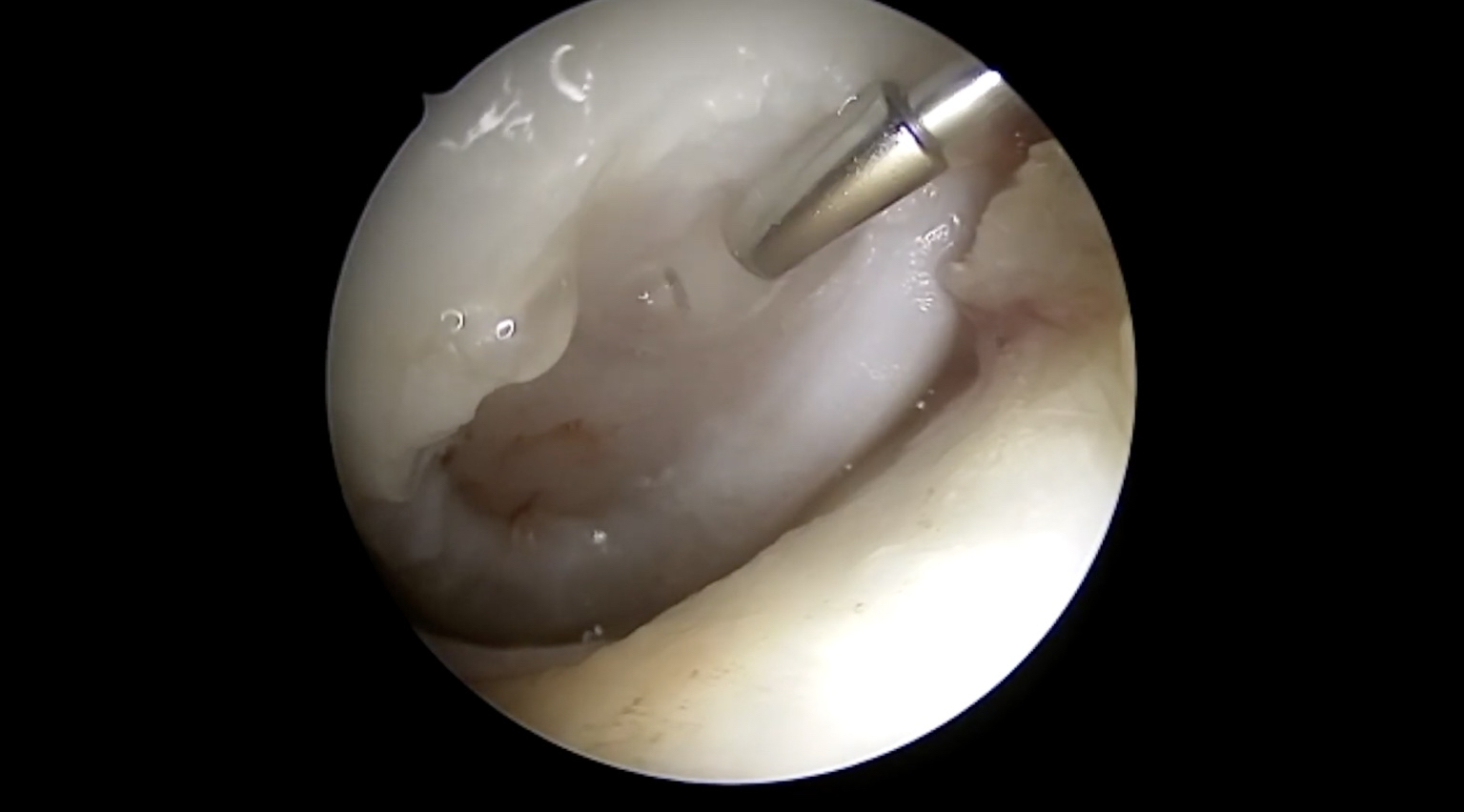
Arthroscopic Implantation
The autologous chondrocytes embedded in the hydrogel are applied directly into the defect together with the crosslinking agent using a dual-chamber syringe. Within approximately 1 to 3 minutes, a stable matrix forms, securing the cells within the defect area. When clinically indicated, the transplantation can be performed arthroscopically.

Recovery and Rehabilitation
Postoperative rehabilitation follows the recommendations of the German Society for Orthopaedics and Trauma Surgery (DGOU) and is largely determined by the location of the cartilage defect within the joint. Following a 48-hour period of rest, gradual mobilization is initiated.For femoral condyle defects, partial weightbearing is recommended until week 6. Two forearm crutches are used until full weight-bearing is achieved.For retropatellar defects, full weight bearing is generally permitted, with a ROM brace and flexion limits determined by defect size and location.
Key Characteristics of Injectable MACT
01
Autologous chondrocytes
No donor tissue; low immunogenic potential
02
Hydrogel matrix
Supports the chondrocyte phenotype and provides favourable conditions for cartilage regeneration
03
Shorter procedure time
In situ cross-linking of the hydrogel within 1-3 minutes; no fixation using sutures or glue required
04
Focal defects
Suitable for focal defects ≥2 cm² [1]
05
Suitable for outpatient treatment
The procedure may be performed on an outpatient basis in appropriate patients [6]
The Procedure Explained Step by Step
This video shows the intraoperative application of Injectable MACT, including defect preparation and in situ gel formation.
Indications
When to Use Our Products
Injectable MACT may be considered for the treatment of symptomatic full-thickness cartilage defects surrounded by healthy adjacent cartilage. In patients with advanced osteoarthritis, biological cartilage repair procedures are generally no longer indicated and alternative treatment options, including joint replacement, should be considered.
Patient-Specific Factors in Treatment Selection
The choice of treatment should be based on an individual clinical assessment. In addition to defect characteristics, treatment selection should take into account several patient-specific factors, including:
Previous procedures on the affected joint
Body weight
Biological age
Physical activity level and sports participation
Comorbidities and risk factors (e.g. smoking, metabolic disorders)
Detailed Indications*
MACT may be considered in patients with the following characteristics:
Symptomatic chondral or osteochondral lesions Grade III-IV (ICRS) - presenting as full-thickness cartilage defects associated with clinically relevant symptoms
Focal cartilage defects of the knee - including defects resulting from trauma, overuse, or osteochondritis dissecans
Defect sizes between 2 and 10 cm² (Scaffold-Based MACT) or ≥2 cm² (Injectable MACT)
Closed epiphyseal growth plates in the knee to be treated
Stable ligamentous structures; existing ligament instability should be addressed in conjunction with MACT.
Intact or functionally sufficient meniscus - partial resection of up to one-third of total meniscal volume is acceptable.
Normal or correctable limb alignment - axis deviations greater than 3° should be corrected in conjunction with MACT
No advanced osteoarthritis - Kellgren-Lawrence Grade ≤2
*For further product-specific information on Scaffold-Based MACT and Injectable MACT, including detailed usage guidance, see the “Healthcare Professional Resources” section.
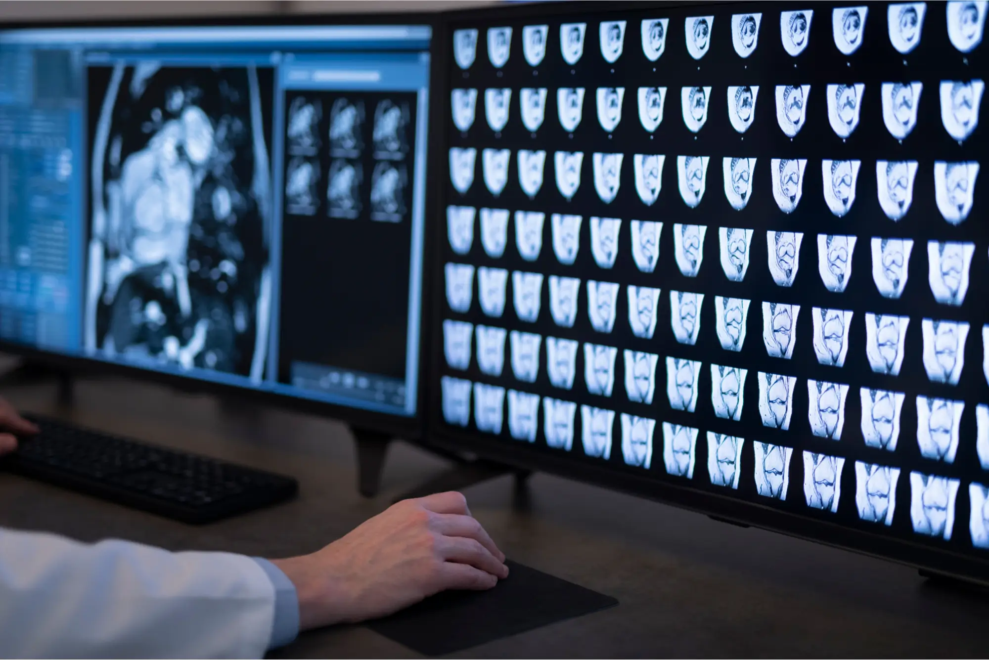

How We Expand Cells
Follow every step of the MACT manufacturing process, from biopsy to final transplant, and learn how controlled manufacturing conditions and continuous quality controls contribute to consistently high transplant quality.
Clinical Outcomes
Available clinical evidence has demonstrated sustained positive outcomes for Injectable MACT, alongside low failure and revision rates.
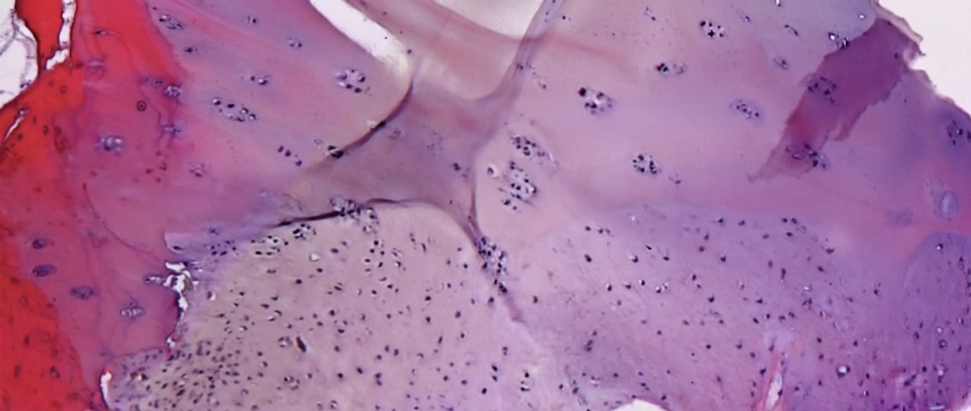
Potential Side Effects
The following side effects may occur with MACT [1]. Additional information is available in the Healthcare Professional Resources section.
Knee pain and swelling
Joint effusion
Joint inflammation or infection
Temporary muscle atrophy during the unloading phase
Temporary stiffness or restricted range of motion
Postoperative thrombosis
Graft failure or insufficient cartilage regeneration
Why Consider Injectable MACT?
CONTACT TETEC
1. TETEC AG. Summary of Product Characteristics (SmPC) NOVOCART® Inject. Reutlingen (DE): TETEC AG; 2026. Revision date: 02/2026. PEI.A.11763.01.1.
2. Niemeyer P, Hanus M, Belickas J, et al. Treatment of large cartilage defects in the knee by hydrogel-based autologous chondrocyte implantation: two-year results of a prospective, multicenter, single-arm phase III trial. Cartilage. 2022;13(1):19476035221085146. doi:10.1177/19476035221085146.
3. Gemeinsamer Bundesausschuss. Beschluss über eine Änderung der Richtlinie Methoden vertragsärztliche Versorgung: Matrixassoziierte autologe Chondrozytenimplantation am Kniegelenk. Berlin (DE): G-BA; 17. Februar 2022.
4. Niemeyer P, Hanus M, Belickas J, et al. Treatment of large cartilage defects in the knee by hydrogel-based autologous chondrocyte implantation: a 5-year follow-up of a prospective, multicenter, single-arm phase III trial. Cartilage. 2025. Epub ahead of print. doi:10.1177/19476035251334737.
5. Richter J, Schuster P. Arthroskopische ACT: die nächste Generation der Knorpelzelltransplantation. Sportärztezeitung. 2016:48–50.
6. Schlumberger M, Schuster P, Bülow HJ, et al. Arthroscopic autologous chondrocyte implantation in the knee with an in situ crosslinking matrix: minimum 4-year clinical results of 15 cases and 1 histological evaluation. Arch Orthop Trauma Surg. 2019;139(11):1607–1615.